
Navicular syndrome — why podotrochlosis is a complex and not a single finding
Navicular bone, navicular bursa and deep digital flexor tendon work as one mechanical system. When this system is chronically overloaded, a multilayered pain process arises in the horse's hoof — with hoof balance and even movement as the central adjusting screws.
Equiterna Redaktion
Veterinarian · Sports medicine
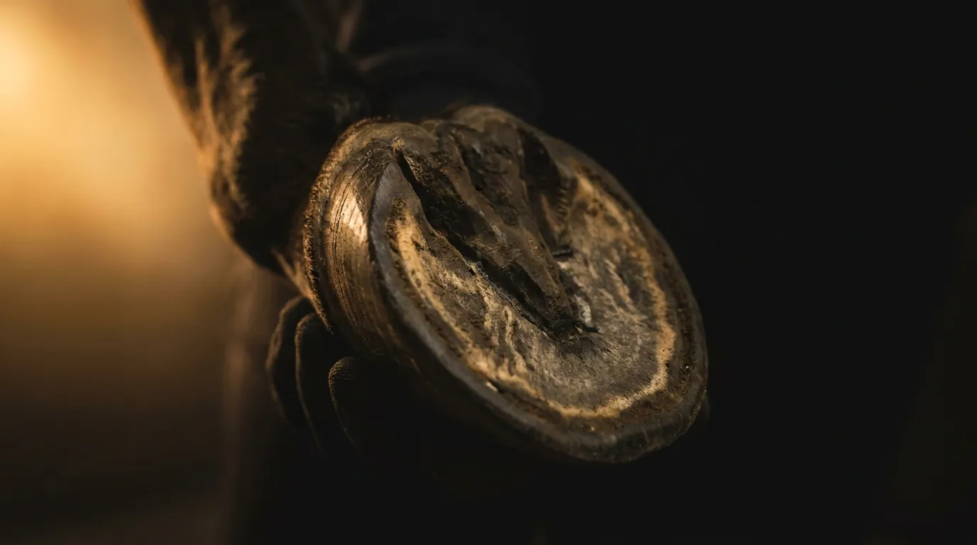
The term navicular region describes no single structure but a functional apparatus deep inside the hoof. Three building blocks engage mechanically with one another here: the navicular bone — a small, transverse bone behind the coffin joint —, the navicular bursa lying beneath it as a gliding cushion, and the deep digital flexor tendon, which runs over the navicular bone like over a pulley before it inserts on the coffin bone. This arrangement gives the apparatus its name: bursa, navicular bone and tendon together form the podotrochlea.
With every footfall the deep digital flexor tendon runs over the gliding surface of the navicular bone and generates high pressure there. As long as load and tissue stay in balance, the system glides with little friction. If this balance tips over a long time, not a single structure begins to suffer but the whole assembly — exactly this is what makes the disease picture so multilayered.
Why podotrochlosis is a complex of chronic changes
The modern term podotrochlosis has deliberately replaced the old word navicular inflammation. It makes clear that not a single finding but a complex of chronic changes is present: remodelling processes on the bone and on the gliding surface of the navicular bone, irritation and thickening of the bursa, as well as damage to the deep digital flexor tendon — from superficial fraying to fibre tears within the tendon. Which of these components stands in the foreground differs from horse to horse, and they often condition one another. That is why today one rather speaks of palmar foot pain: a collective term for pain in the back part of the hoof with several possible sources.
Typical signs — quiet and bilateral
Palmar foot pain rarely announces itself dramatically. Often a stiff, shortened foreleg action is the first to be noticed: the horse steps shorter in front, seems stiff when trotting off and noticeably seeks soft ground, because hard, uneven footing increases the pressure in the navicular region. On tight turns or on the circle the lameness can intensify. Because both front hooves are often similarly affected, the clear limping on one leg is missing — instead the complaints show as a general reluctance, as stumbling or as an altered, cautious footfall. Some horses place the painful foot conspicuously forward in the box to relieve the deep digital flexor tendon.
Precisely this quietness is treacherous: what is dismissed as declining rideability or an unclean canter can be the first hint of a chronic process inside the hoof. The classification of such signs belongs in veterinary hands.
Hoof balance, shoeing and even movement
The mechanics of the navicular region hang directly on the shape and position of the hoof. A long toe with low, underrun heel lengthens the lever during breakover and increases the pull of the deep digital flexor tendon over the navicular bone — exactly where the system is already most heavily loaded. A balanced hoof with correct toe-heel axis and a clean, regular breakover therefore belong to the foundation of every management. Hoof care and, where appropriate, shoeing are no cosmetic step here, but directly mechanically effective.
Equally important is even, measured movement on suitable ground. Regular, controlled load keeps the navicular apparatus in function and promotes circulation; abrupt load peaks, hard or deeply uneven footing and tight, fast turns act against it. Which ground, which hoof shape and which amount of movement suit an individual horse cannot be fixed in a blanket manner — that follows from the concrete findings.
“The navicular region does not punish the one wrong step, but the permanently unfavourable lever. Whoever sets the balance of the hoof correctly takes a part of the load off the tendon over the navicular bone.”
Classification: an insidious, bilateral forehand lameness belongs in veterinary clarification. Only the examination with flexion tests, diagnostic anaesthesia and imaging — radiography for the navicular bone, MRI for bursa and deep digital flexor tendon — can distinguish the structures involved. This article explains the disease picture and the importance of hoof balance and movement; it makes no diagnosis, gives no therapy instruction and no promise of cure.
Sources
Usage note
This content is for information and education. It does not replace veterinary diagnosis or treatment. If your horse shows signs of illness, consult a veterinarian.
Keep reading
Summer eczema — why the skin is rarely where it starts
The reaction to the midge bite is the visible part. What lies beneath it decides the course.
HealthGrowing old without looking old — what really counts in the senior
Many complaints that pass for age-related are not. A look at what can be influenced.
HealthWhen the coat whispers — what the coat change reveals about the metabolism
Twice a year the horse rebuilds its entire coat. What happens in the metabolism while it does — and which signs can be read.